 Pagal World
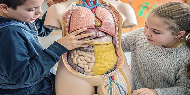
The two divisions of the digestive system are the alimentary tube and the accessory organs. The alimentary tube extends from the mouth to the anus. It consists of the oral cavity, pharynx, esophagus, stomach, small intestine, and large intestine. Digestion takes place within the oral cavity, stomach, and small intestine; most absorption of nutrients takes place in the small intestine. Indigestible material, primarily cellulose, is eliminated by the large intestine. The accessory organs of digestion are the teeth, tongue, salivary glands, liver, gallbladder, and pancreas. Digestion does not take place within these organs, but each contributes something to the digestive process.
ORAL CAVITY
Food enters the oral cavity (or buckle cavity) by way of the mouth. The boundaries of the oral cavity are the hard and soft palates superiorly; the cheeks laterally; and the floor of the mouth inferiorly. Within the oral cavity are the teeth and tongue and the openings of the ducts of the salivary glands.
SALIVARY GLANDS
The digestive secretion in the oral cavity is saliva, produced by three pairs of salivary glands. The parotid glands are just below and in front of the ears. The submandibular (also called sub maxillary) glands are at the posterior corners of the mandible, and the sublingual glands are below the floor of the mouth. Each gland has at least one duct that takes saliva to the oral cavity. Secretion of saliva is continuous, but the amount varies in different situations. The presence of food (or anything else) in the mouth increases saliva secretion. This is a parasympathetic response mediated by the facial and glossopharyngeal nerves. The sight or smell of food also increases secretion of saliva. Sympathetic stimulation in stress situations decreases secretion, making the mouth dry and swallowing difficult.
ESOPHAGUS
The esophagus is a muscular tube that takes food from the pharynx to the stomach; no digestion takes place here. Peristalsis of the esophagus propels food in one direction and ensures that food gets to the stomach even if the body is horizontal or upside down. At the junction with the stomach, the lumen (cavity) of the esophagus is surrounded by the lower esophageal sphincter (LES or cardiac sphincter), a circular smooth muscle. The LES relaxes to permit food to enter the stomach, then contracts to prevent the backup of stomach contents. If the LES does not close completely, gastric juice may splash up into the esophagus; this is a painful condition we call heartburn, or gastro esophageal reflux disease (GERD). Most people experience heartburn once in a while, and it is merely uncomfortable, but chronic GERD is more serious. The lining of the esophagus cannot withstand the corrosive action of gastric acid and will be damaged, perhaps resulting in bleeding or even perforation. Medications are available to treat this condition.
STRUCTURAL LAYERS OF THE ALIMENTARY TUBE
Before we continue with our discussion of the organs of digestion, we will first examine the typical structure of the alimentary tube. When viewed in cross-section, the alimentary tube has four layers (Fig. 16–4): the mucosa, sub mucosa, external muscle layer, and serosa. Each layer has a specific structure, and its functions contribute to the functioning of the organs of which it is a part.
SUBMUCOSA
The sub mucosa is made of areolar connective tissue with many blood vessels and lymphatic vessels. Many millions of nerve fibers are also present, part of what is called the enteric nervous system, or the “brain of the gut,” which extends the entire length of the alimentary tube. The nerve networks in the sub mucosa are called Meissen’s plexus (or sub mucosal plexus), and they innervate the mucosa to regulate secretions. Parasympathetic impulses increase secretions, whereas sympathetic impulses decrease secretions. Sensory neurons are also present to the smooth muscle (a stretched or cramping gut is painful), as are motor neurons to blood vessels, to regulate vessel diameter and blood flow.
EXTERNAL MUSCLE LAYER
The external muscle layer typically contains two layers of smooth muscle: an inner, circular layer and an outer, longitudinal layer. Variations from the typical do occur, however. In the esophagus, this layer is striated muscle in the upper third, which gradually changes to smooth muscle in the lower portions. The stomach has three layers of smooth muscle, rather than two.
SUMMARY
The processes of the digestion of food and the absorption of nutrients enable the body to use complex food molecules for many purposes. Much of the food we eat literally becomes part of us. The body synthesizes proteins and lipids for the growth and repair of tissues and produces enzymes to catalyze all of the reactions that contribute to homeostasis. Some of our food provides the energy required for growth, repair, movement, sensation, and thinking. In the next chapter we will discuss the chemical basis of energy production from food and consider the relationship of energy production to the maintenance of body temperature